- Home
- About Us
- Meet Our Team
- Patient Forms
- FAQs
- Services
- Hearing Evaluations And Testing
- Hearing Aid Fitting And Repair
- Custom Ear Molds And Ear Plugs
- Tinnitus Evaluation
- Hearing Aids
- Types of Hearing Aids
- Smart Hearing Aids
- Hearing Aids We Carry
- Caring for Your Hearing Aids
- Hearing Loss and Brain Health
- Hearing Health
- What to Expect at Your First Visit
- Types of Hearing Loss
- How The Ear Works
- Aftercare
- Online Hearing Test
- Blog
- Contact Us
- New Alexandria
- Greensburg
- Ligonier
- Monroeville
- Shop
- Review Us
- New Alexandria
- Greensburg
- Book an Appointment
About Us
Services
Hearing Aids
Hearing Health
Contact Us
Review Us

Normal Hearing Function
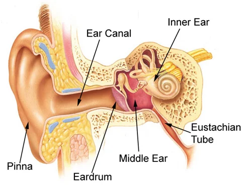
- Sound is transmitted through the air as sound waves and are gathered by the outer ear and sent down the ear canal to the eardrum.
- The sound waves cause the eardrum to vibrate, which sets the three tiny bones in the middle ear into motion.
- The motion of the three bones causes the fluid in the inner ear, or cochlea, to move.
- The movement of the fluid in the inner ear causes the hair cells in the cochlea to bend. The hair cells change the movement into electrical impulses.
- These electrical impulses are transmitted to the auditory nerve and up to the brain, where they are interpreted as sound.
The Outer Ear
The part of the outer ear that we see is called the pinna, or auricle. The pinna, with its grooves and ridges, provides a natural volume boost for sounds in the 2000 to 3000 Hz frequency range, where we perceive many consonant sounds of speech.
The ear canal, also called the external auditory meatus, is the other important outer ear landmark. This part of the ear is lined with only a few layers of skin and fine hair, with many veins traveling all around it. This means that there is an abundant flow of blood to the ear canal. Earwax (cerumen) accumulates in the ear canal and serves as a protective barrier to the skin from bacteria and moisture. Earwax is normal, unless it completely blocks the ear canal.
The Middle Ear
The eardrum, or tympanic membrane (TM), is the dividing structure between the outer and middle ear. Although it is an extremely thin membrane, the eardrum is made up of three layers to increase its strength.
The ossicles are the three tiny bones of the middle ear located directly behind the tympanic membrane. These are the three tiny bones (smallest in the human body) in the middle ear: malleus (hammer), incus (anvil,) and stapes (stirrup) and their job is to further amplify the sound. These three bones form a connected chain in the middle ear. The malleus is embedded in the innermost layer of the tympanic membrane, and the stapes is connected to a membranous window of the inner ear, called the oval window. The ossicles translate mechanical vibrations received at the eardrum into the inner ear.
The Eustachian tube is the middle ear’s air pressure equalizing system. The middle ear is encased in bone and does not associate with outside air except through the Eustachian tube. This tubular structure is normally closed, but it can be involuntarily opened by swallowing, yawning, or chewing. It can also be intentionally opened to equalize pressure in the ears, such as when flying in an airplane. When this happens, you might hear a soft popping sound.
The Inner Ear
The inner ear is an organ located deep within the temporal bone, which is the bone of the skull on both sides of the head above the outer ear. The inner ear has two main structures: the semicircular canals and the cochlea.
The semicircular canals do not contribute to hearing, but assist in maintaining balance as we move. The cochlea is the hearing organ of the inner ear, which is a fluid-filled structure that looks like a snail’s shell. The cochlea changes the mechanical vibrations from the eardrum and the ossicles into a sequence of electrical impulses. Sensory cells, called hair cells, bend in the cochlea as the fluid is disrupted by the mechanical vibrations. This bending of the hair cells causes electrical signals to be sent to the brain by way of the auditory nerve. The cochlea is arranged by frequency, much like a piano, and encodes sounds from 20Hz (low pitch) to 20,000Hz (high pitch) in humans.
